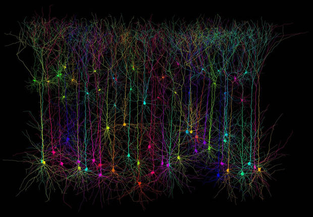
Michael Hausser and Hermann Cuntz, UCL
Pyramidal neurons are located in the forebrain of mammals and associated with the process cognitive function. This is a computer-simulated picture of how they would look in real life.
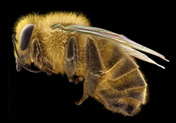
David McCarthy and Annie Cavanagh
False-colored scanning electron micrograph of a honeybee.
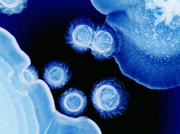
Derren Ready, Eastman Dental Institute
If this color-enhanced image of periodontal bacteria doesn't convince you to brush twice a day AND visit a dentist regularly, then you're simply hopeless.
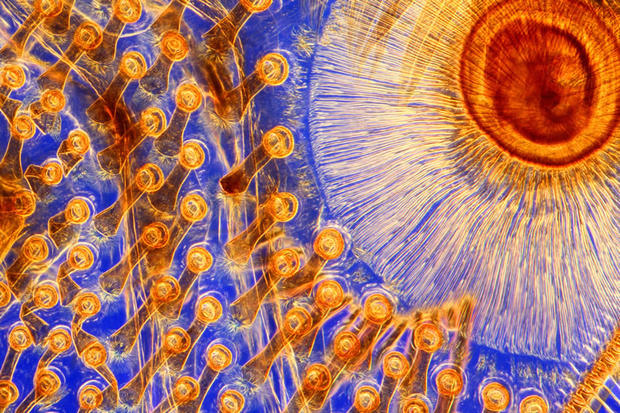
Spike Walker
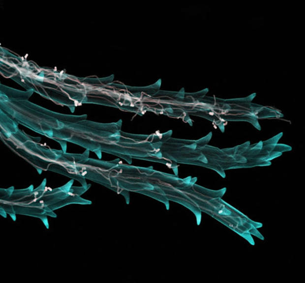
Rows of suckers on the foreleg of a great diving beetle - the largest freshwater beetles in the United Kingdom. This image was produced by passing light through colored filters.
Spike Walker
The base of a silkworm caterpillar's proleg. These are stubby structures that grow from the underside of the caterpillar's abdomen but disappear as the caterpillar grows.
Kara Cerveny, Steve Wilson's Lab, UCL
Retina from the eye of a three-day-old zebrafish. The retina is viewed here from the front.
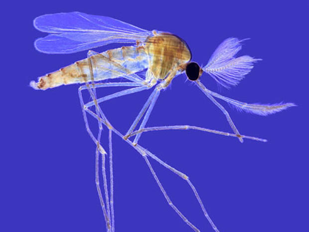
Spike Walker
An adult male mosquito never looked so good - especially considering how sample used for this image was taken from a microscope slide created in the middle of the 20th century.
Anne Weston, London Research Institute, Cancer Research UK
Underside of a band-aid used to treat a razor blade cut.
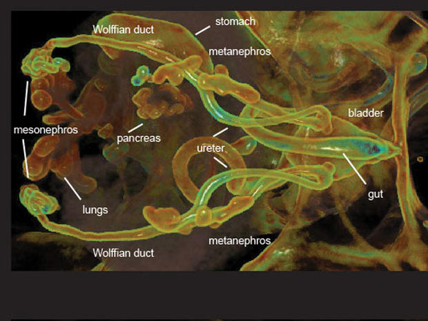
Ian Smyth, Monash University
3-D animation of the developing organs in a mouse embryo. The picture was created from optical projection tomography data.
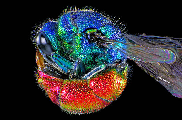
Spike Walker
Adult ruby-tailed wasp curled into a ball. Two electronic flashes added extra imaging to highlight the wasp's colors.
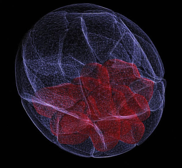
Agnieszka Jedrusik and Magdalena Zernicka-Goetz, Gurden Institute, Cambridge
3D reconstruction of a mouse embryo at the blastocyst stage.
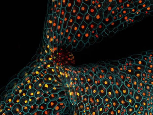
Fernan Federici, University of Cambridge and Lionel Dupuy, Scottish Crop Research Institute
Fluorescent proteins in the stem of a thale cress seedling. It also was the first plant to have its entire genome sequenced.
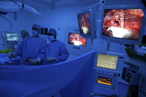
David Bishop, UCK Medical School
Surgeons using a specialized camera to perform a laparoscopy, an operation in which small, thin instruments get placed into the patient's abdomen via small incisions.
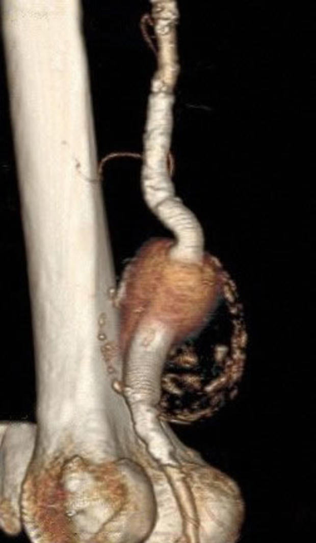
Arindam Chaudhuri
A view of a popliteal aneurysm taken from CT scans of an 84-year-old man undergoing treatment. You can see the aneurysm in the center frame, with its reddish-blue appearance and eggshell-like boundary.
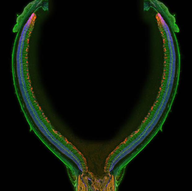
Freya Mowat, UCL
Retina from a one-month-old juvenile mouse.The image was created by 'stitching' and then aligning six smaller images.
Anna Gordon, National Institute of Agricultural Biology, and Fernan Federici, University of Cambridge
Wheat stigma hairs (blue) infected with ergot fungus (light pink).
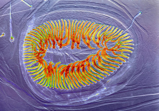
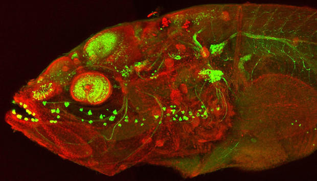
Monica Folgueira, Steve Wilson's Lab, UCL
Cavefish embryo at around five days post-fertilization.
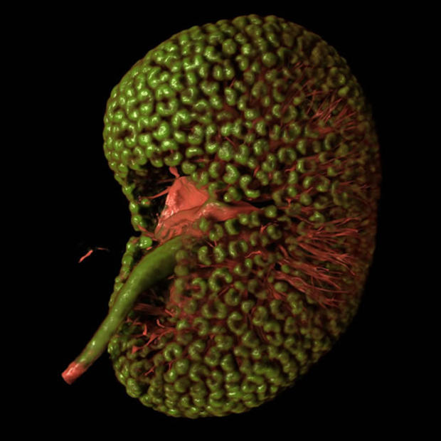
Bob Kao and Kieran Short, Monash University
Fetal mouse kidney at embryonic day 16.
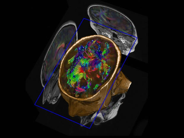
Nuada Medical Specialist Imaging
The neuronal tracts in the brain of an adult male.
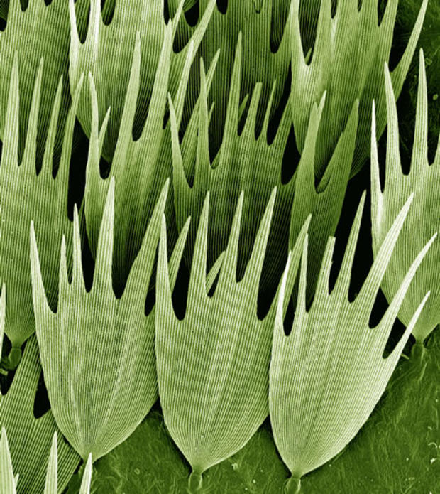
Kevin Mackenzie, University of Aberdeen
Scanning electron micrograph of the scales on the wing of a Madagascan moon moth.
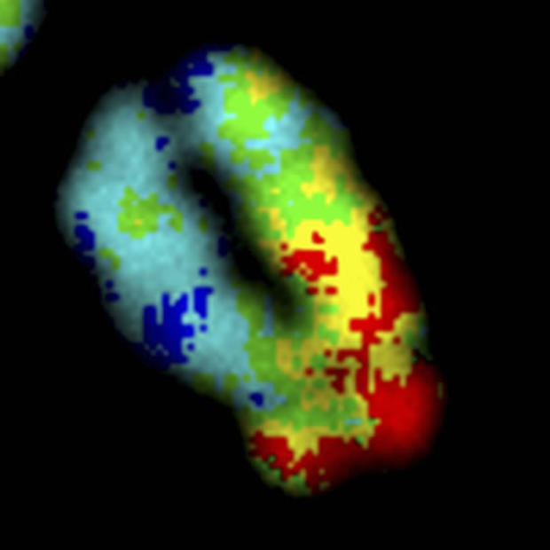
David Lleres, University of Dundee
Image of a human chromosome.