Microscopic beauty
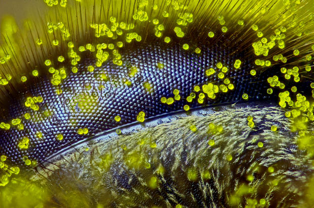
First Prize in the 41st Annual Nikon Small World Photomicrography Competition was awarded to Australian Ralph Claus Grimm for capturing an amazing close-up of the eye of a honey bee covered in dandelion pollen.
Grimm, a high school teacher from Queensland, self-taught microscopist and former bee keeper, was recognized along with more than 80 other international winners. He hopes his image can serve as a voice for this endangered insect that plays such a critical function in pollinating the world's crops.
Read on to see more winning images from the competition.
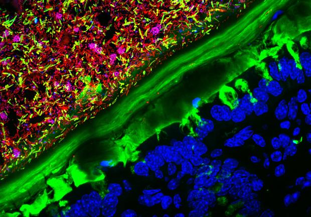
2nd Place
An image of the colon of a mouse that was born germ-free, or completely sterile of microbes. The mouse was colonized with a human microbiota.
Photographed by Kristen Earle, Gabriel Billings, KC Huang & Justin Sonnenburg, Stanford, California
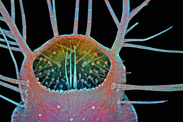
3rd Place
This image shows an entrance to the trap (or bladder) of a Humped Bladderwort (Urticulatia gibba), a carnivorous freshwater plant.
Photographed by Dr. Igor Siwanowicz, Ashburn, Virginia
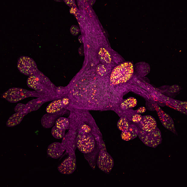
4th Place
A photo of a mini-organ, known as an organoid, grown from human mammary gland tissue in a lab.
Photographed by Daniel H. Miller & Ethan S. Sokol, Cambridge, Massachusetts
5th Place
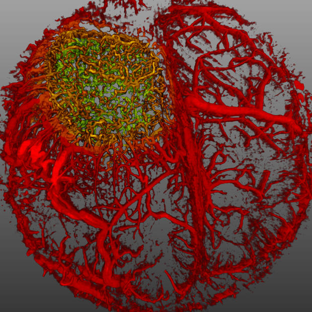
Live imaging of perfused vasculature in a mouse brain with glioblastoma.
Photographed by Dr. Giorgio Seano & Dr. Rakesh K. Jain, Harvard Medical School, Boston
6th Place
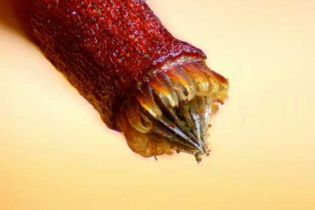
Spore capsule of a moss (Bryum sp.)
Photographed by Henri Koskinen, Helsinki, Finland
7th Place
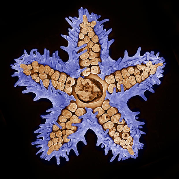
A starfish photographed using confocal microscopy.
Photographed by Evan Darling, Memorial Sloan Kettering Cancer Center, New York
8th Place
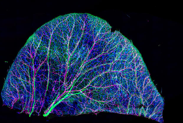
An image of nerves and blood vessels in a mouse ear skin.
Photographed by Dr. Tomoko, Yamazaki National Institutes of Health (NIH), Bethesda, Maryland
9th Place
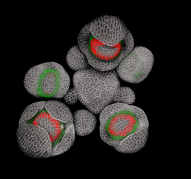
Young buds of Arabidopsis (a flowering plant).
Dr. Nathanael Prunet, Pasadena, California
10th Place
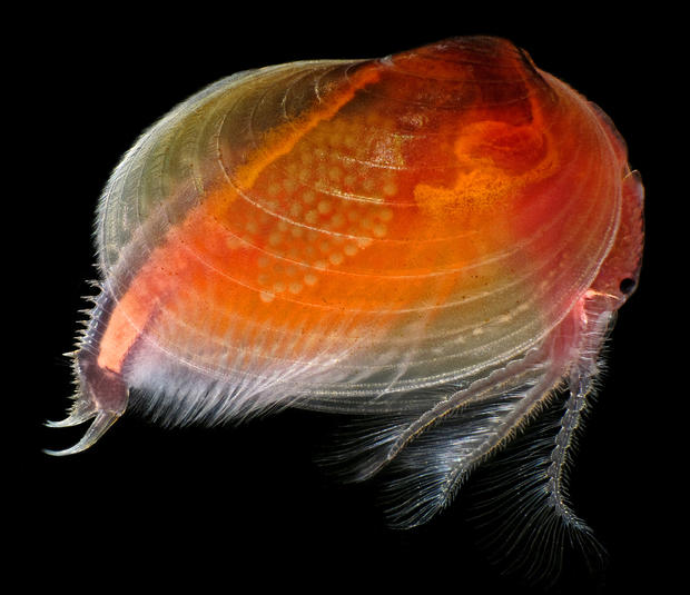
A live clam shrimp (Cyzicus mexicanus).
Photographed by Ian Gardiner, Calgary, Canada
11th Place
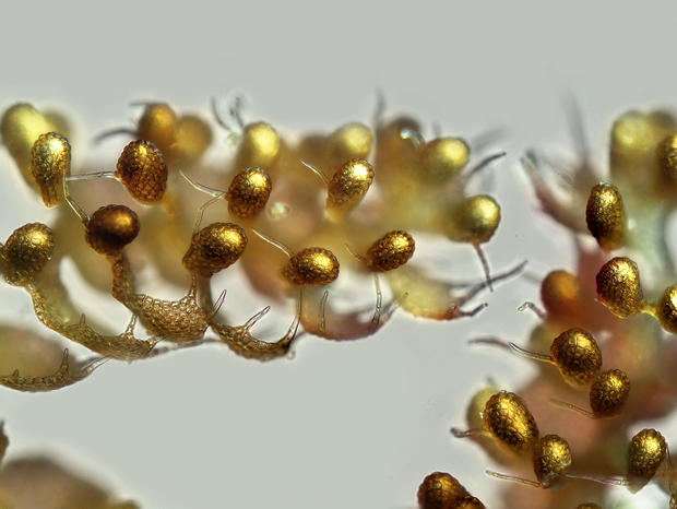
Fern sorus at varying levels of maturity.
Photographed by Rogelio Moreno Gill, Panama
12th Place
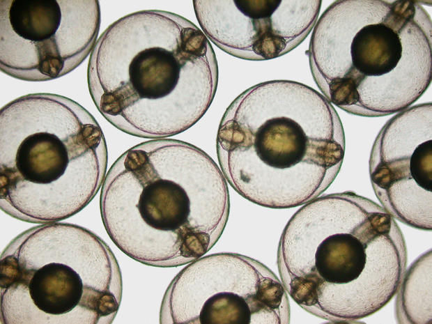
Developing sea mullet (Mugil cephalus) embryos.
Hannah Sheppard-Brennand, Sydney, Australia
13th Place
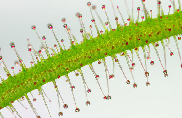
This image shows tentacles of a carnivorous plant (Drosera sp.).
Photographed by Jose R Almodovar, University of Puerto Rico
14th Place
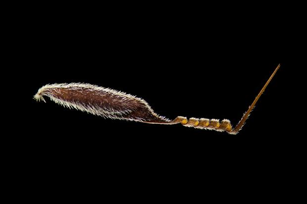
An Australian grass (Austrostipa nodosa) seed.
Photographed by Viktor Sykora, Prague, Czech Republic
15th Place
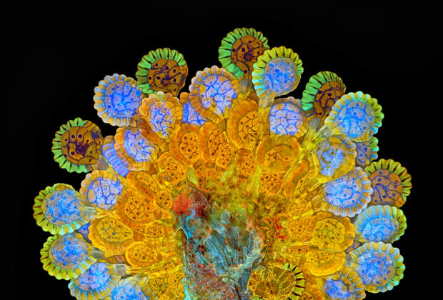
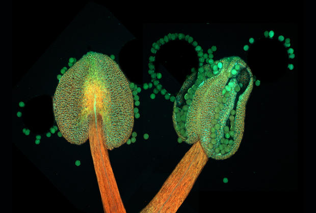
The anthers of a flowering plant (Arabidopsis thaliana).
Photographed by Dr. Heiti Paves, Tallinn, Estonia
16th Place
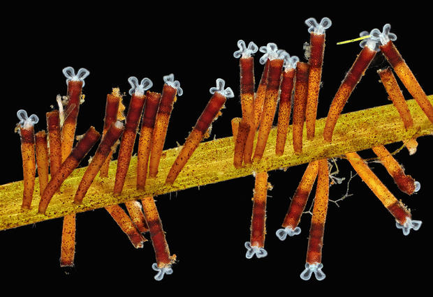
An image of feeding rotifers (Floscularia ringens).
Photographed by Charles B. Krebs, Issaquah, Washington
17th Place
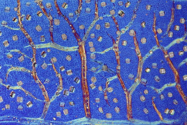
A black witch-hazel (Trichodactylus crinitus) leaf producing crystals to defend against herbivores.
Photographed by Dr. David Maitland, Feltwell, UK
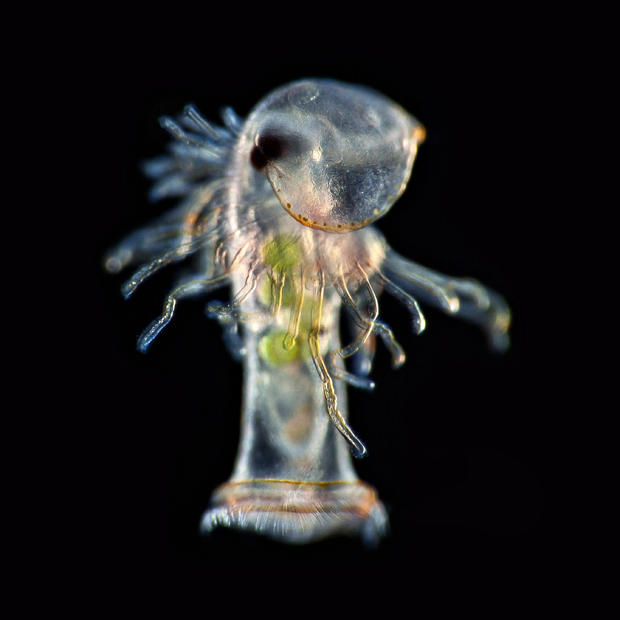
19th Place
Planktonic larva of a horseshoe worm (phoronid).
Photographed by Dr. Richard R. Kirby, UK
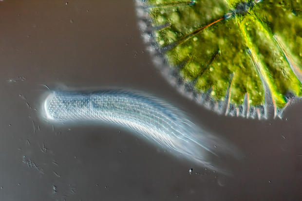
18th Place
A hairyback worm (Chaetonotus sp.) and algae (Micrasterias sp.).
Photographed by Roland Gross, Gruenen, Switzerland
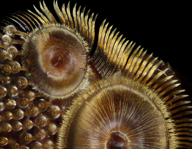
20th Place
The suction cups on a diving beetle (Dytiscus sp.) foreleg.
Photographed by Frank Reiser, Garden City, New York
Honorable Mention
A liverwort (Lepidolaena taylorii) plant with modified leaves (water sacs), which are often home to aquatic microorganisms such as rotifers.
Photographed by Susan Tremblay, Berkeley, California
Honorable Mention
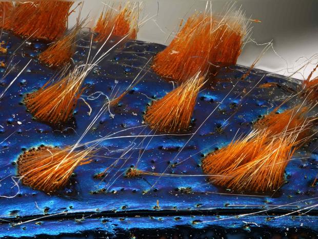
A detail of a jewel beetle (Coleoptera Buprestidae).
Photographed by Dr. Luca Toledano, Verona, Italy
Honorable Mention
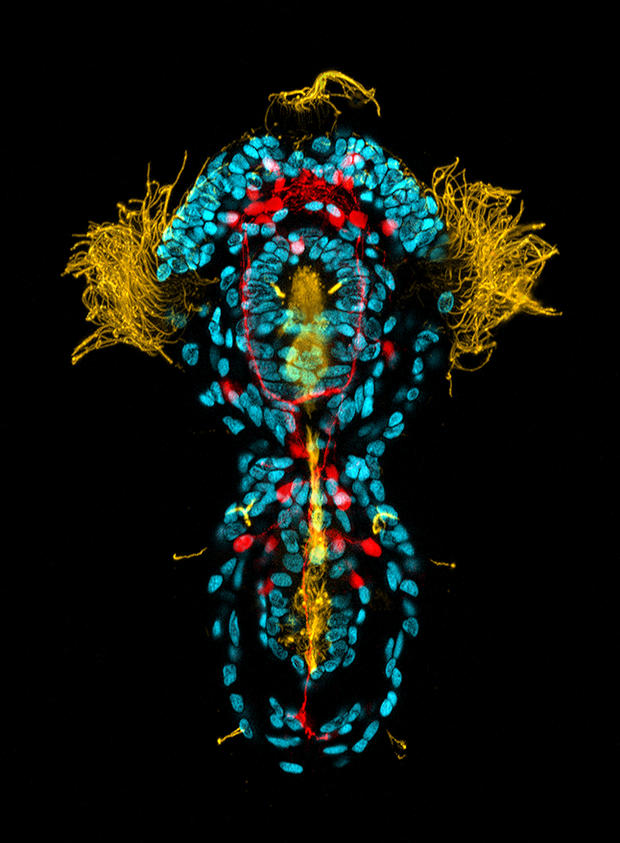
A three-day old Peanut worm (Sipuncula), serotonin in the nervous system.
Photographed by Dr. Michael J. Boyle, Fort Pierce, Florida
Honorable Mention
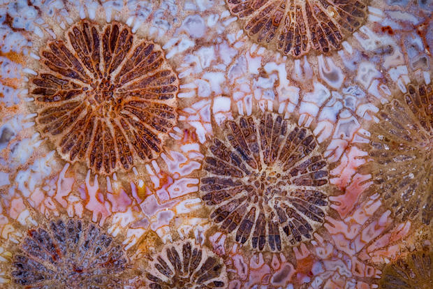
A red fossil coral slab.
Photographed by Norm Baker, Baltimore, Maryland
Honorable Mention
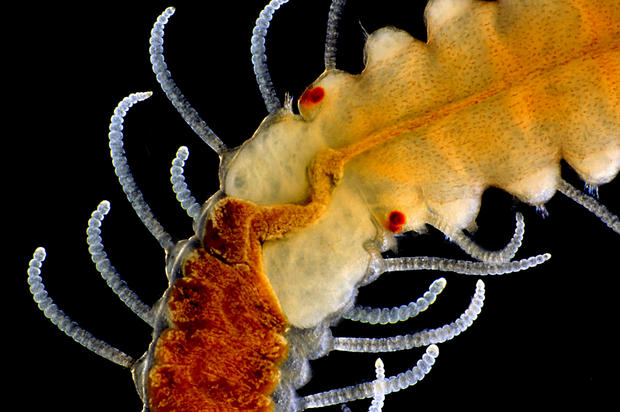
An adult marine worm (Autolytus).
Photographed by Mike Crutchley, Wales, UK
