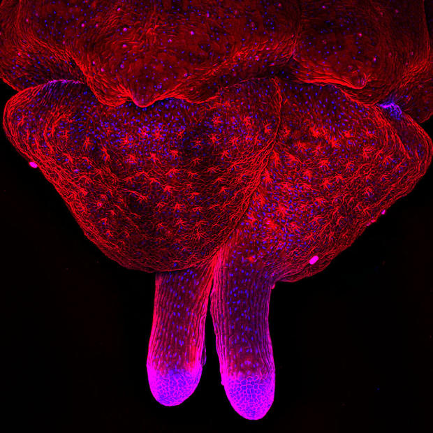
Minuscule beauty
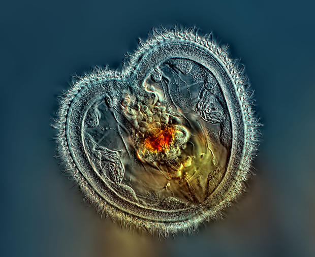
First Prize in the 40th Annual Nikon Small World Photomicrography Competition was awarded to Rogelio Moreno of Panama for capturing this image of a rarely seen image of a rotifer's open-mouthed interior and heart-shaped corona.
Moreno, a self-taught microscopist, was recognized along with more than 80 other international winners.
Read on to see more winning images from the competition.
Second Place
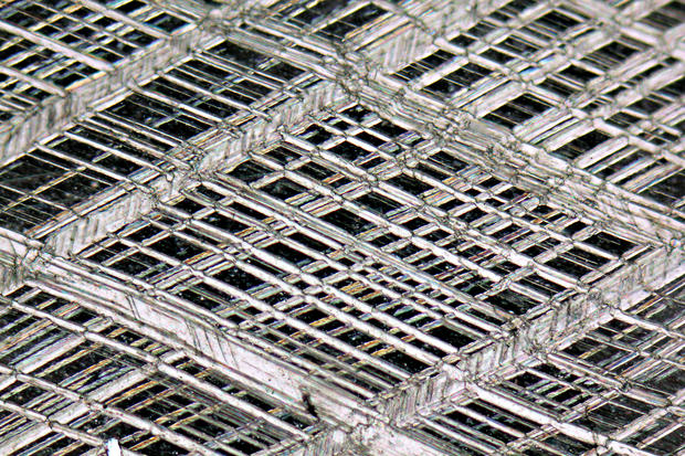
Rhombohedral cleavage in calcite crystal.
Photographed by Alessandro Da Mommio, Pisa, Italy
Third Place
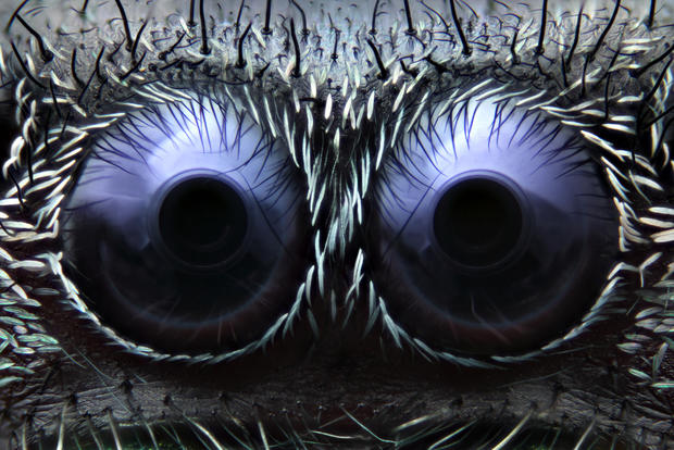
Jumping spider eyes, reflected light.
Photographed by Noah Fram-Schwartz, Greenwich, Connecticut
Fourth Place
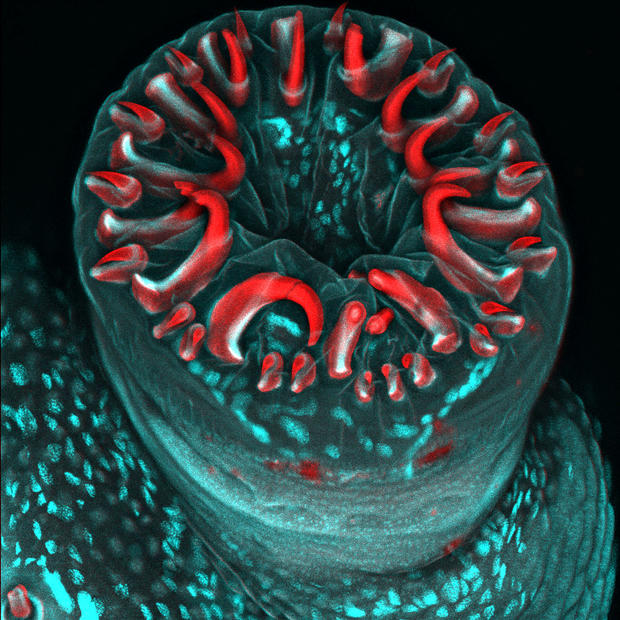
A caterpillar proleg with circle of gripping hooks.
Photographed by Karin Panser, Vienna, Austria
Fifth Place
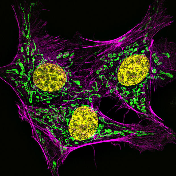
Bovine pulmonary artery endothelial cells stained for actin (pink), mitochondria (green) and DNA (yellow).
Photographed by Muthugapatti K. Kandasamy, Athens, Georgia
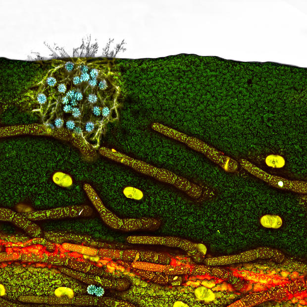
Sixth Place
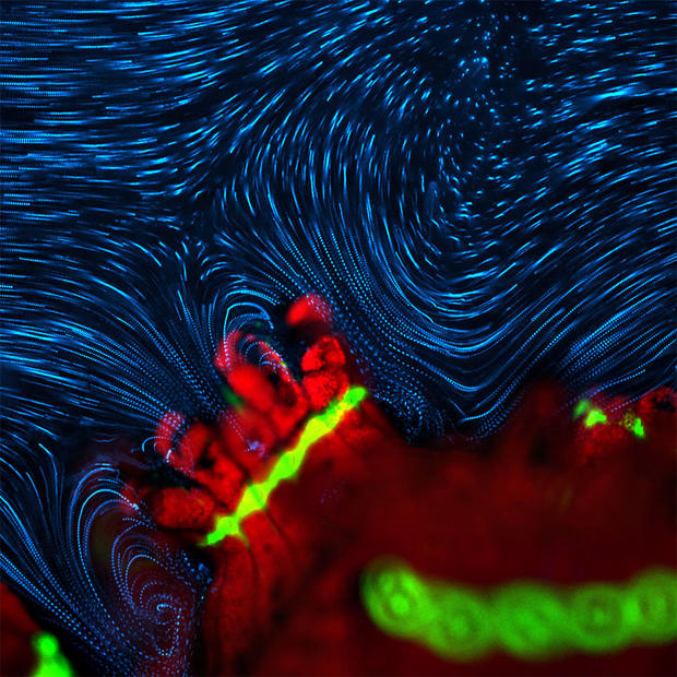
Active fluid flow around coral polyp.
Photographed by Dr. Douglas Brumley, Cambridge, Massachusetts
Seventh Place
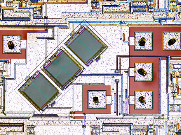
Circuitry in a DVD reader-cross-polarized microscopy.
Photographed by Dennis Hinks, Cleveland, Ohio
Eighth Place
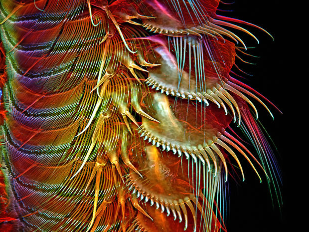
Appendages of a common brine shrimp.
Photographed by Dr. Igor Robert Siwanowicz, Ashburn, Virginia
Ninth Place
Parsley ovary fixed and stained to show lectins (red) and nuclei (blue).
Photographed by Meritxell Vendrell, Barcelona, Spain
Tenth Place
A daisy petal with fungal infection and pollen grains.
Photographed by Dr. Paul Joseph Rigby, Crawley, Western Australia, Australia
11th Place
A house cricket's tongue.
Photographed by Stefano Barone, Cremona, Italy
Twelfth Place
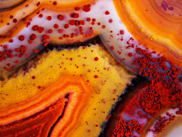
A Montana dryhead agate, unpolished.
Photographed by Douglas Moore, Stevens Point, Wisconsin
Thirteenth Place
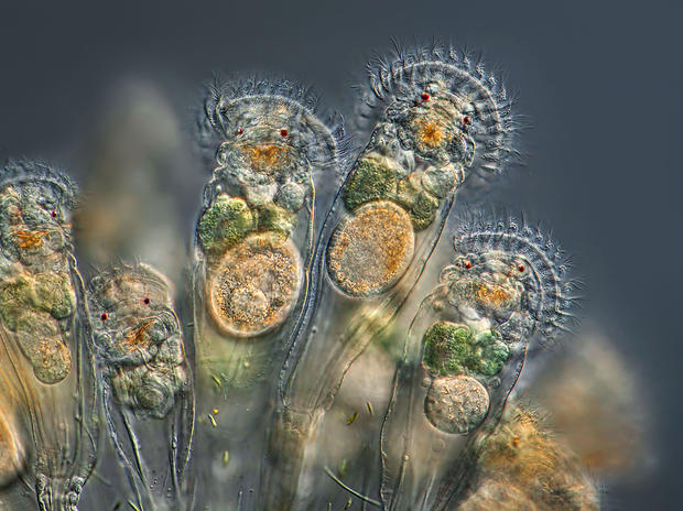
A rotifer actively feeding.
Photographed by Charles Krebs, Issaquah, Washington
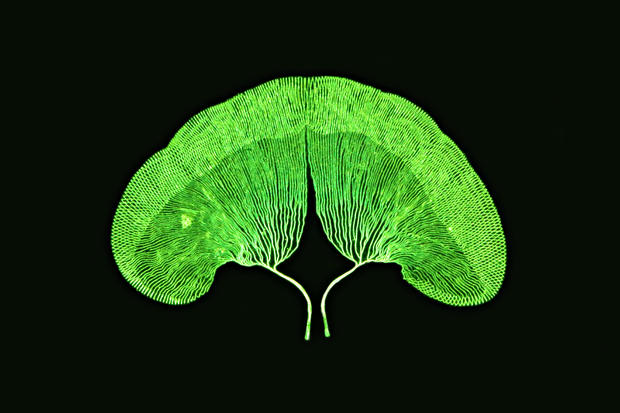
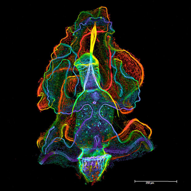
Fourteenth Place
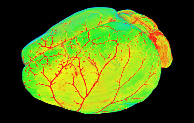
A mouse brain.
Photographed by Dr. Ali Erturk, Munich, Germany
Fifteenth Place
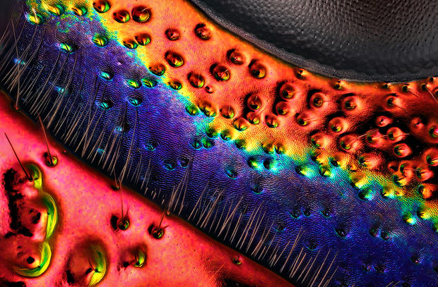
A jewel beetle near the eye.
Photographed by Charles Krebs, Issaquah, Washington
Sixteenth Place
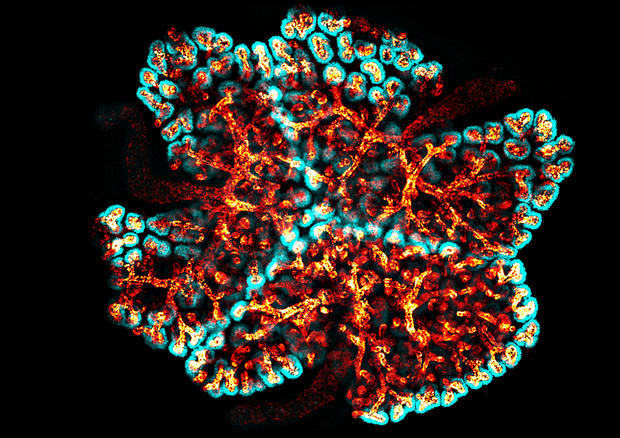
Three transgenic kidneys cultured together, showing colliding branching collecting duct systems.
Photographed by Dr. Nils Lindstrom, Edinburgh, Scotland
Seventeenth Place
Micro algae.
Photographed by Rogelio Moreno, Panama, Panama
Eighteenth Place
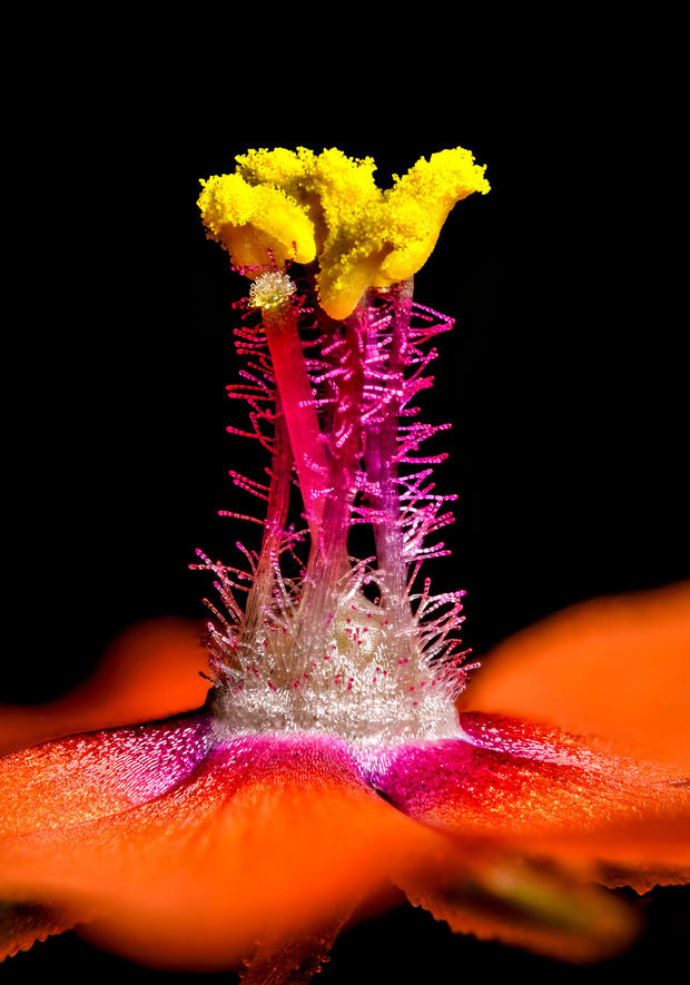
A scarlet pimpernel.
Photographed by Jens H. Petersen, Ebeltoft, Denmark
Ninteenth Place
Larval stage of the acorn worm, showing cell borders, muscles and apical eye spots.
Photographed by Dr. Sabrina Kaul, Vienna, Austria
Twentieth Place
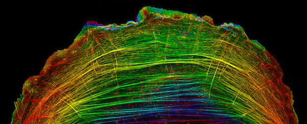
A crawling bone cancer cell.
Photographed by Dr. Dylan T. Burnette, Nashville, Tennessee
Honorable Mention
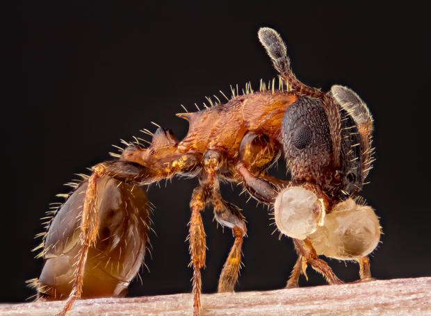
An ant carrying its larva.
Photographed by Geir Drange, Asker, Norway
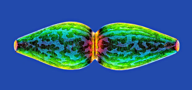
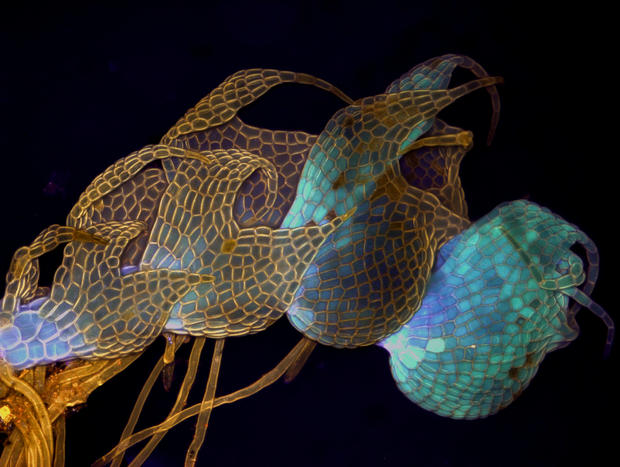
Honorable Mention
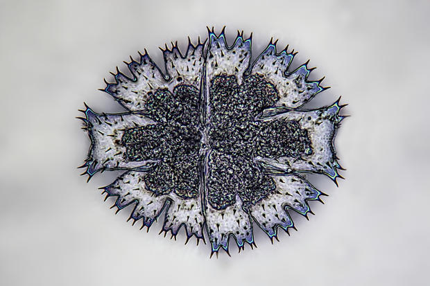
Living Micrasterias.
Photographed by Frank Fox, Konz,Germany
Honorable Mention
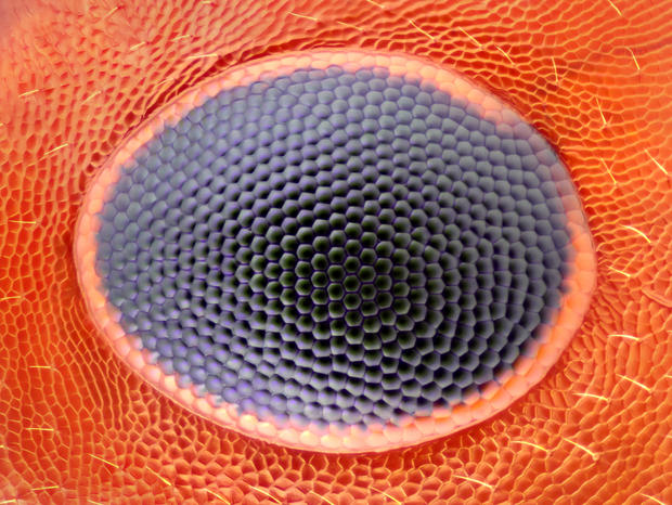
An ant eye.
Photographed by Noah Fram-Schwartz, Greenwich, Connecticut
Honorable Mention
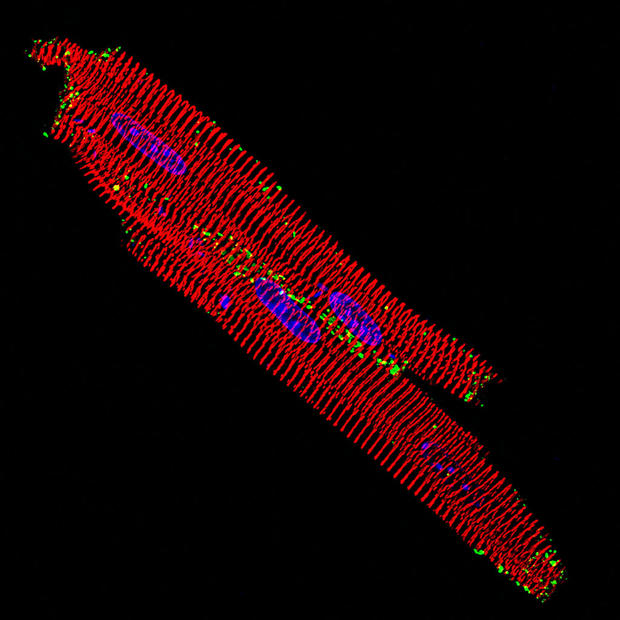
Isolated heart muscles of a mouse.
Photographed by Dr. William James Hatton, Sydney, Australia
Honorable Mention
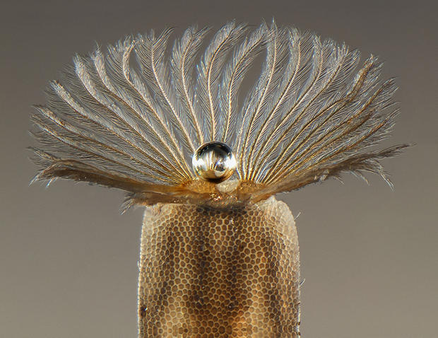
An air pearl in the middle of larva.
Photographed by Fabrice Parals, Caen, France
Honorable Mention
A leafy liverwort.
Photographed by MagdalenaTurzanska, Wroclaw, Poland