Microscopic images show artistry of cellular life
These photos are part of the exhibit “Life: Magnified,” cosponsored by the National Institute of General Medical Sciences, the American Society for Cell Biology and the Metropolitan Washington Airports Authority’s Arts Program, on view through November 2014 at Washington Dulles International Airport.
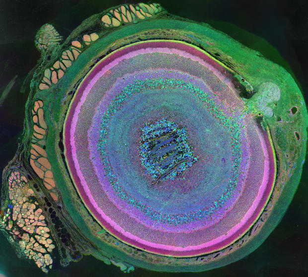
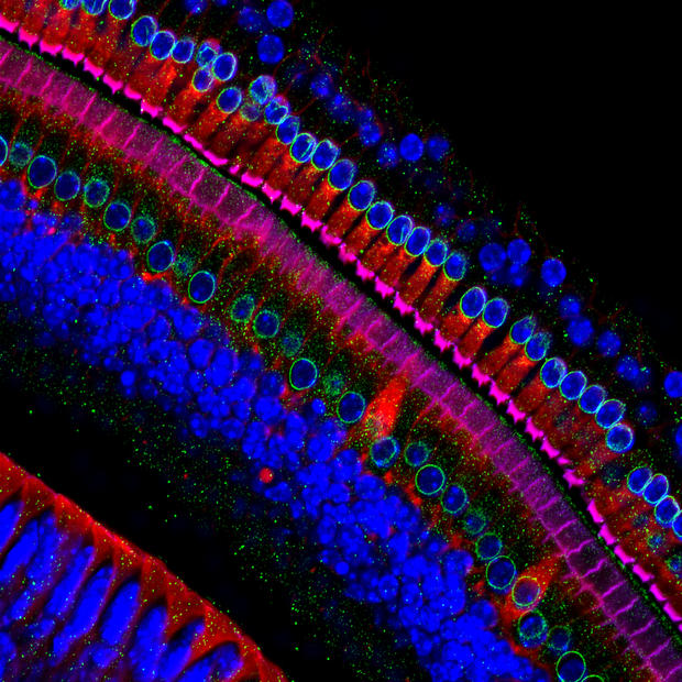
This image of cells from a mouse eye illustrates the complexity of a mammalian eye, which is made up of at least 70 different cell types. Each color in this picture represents a different type, which include the many layers of the retina and peach-colored muscle cells to the left.
Yeast cell is born
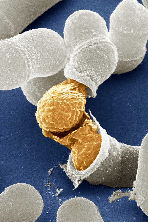
Yeast -- a microorganism found in foods such as beer and bread, as well as in the human body -- can reproduce sexually. A mother and father cell fuse together and create one large cell that results in four offsprings. Here, one offspring emerges.
Mouth of a lone start tick
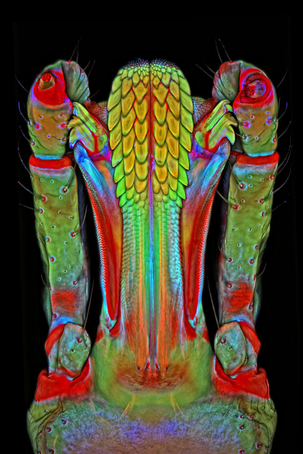
The center of the mouth (yellow) is covered by sharp points, which helps the tick stay secure while feeding on its host.
Immune system devours anthrax bacterium
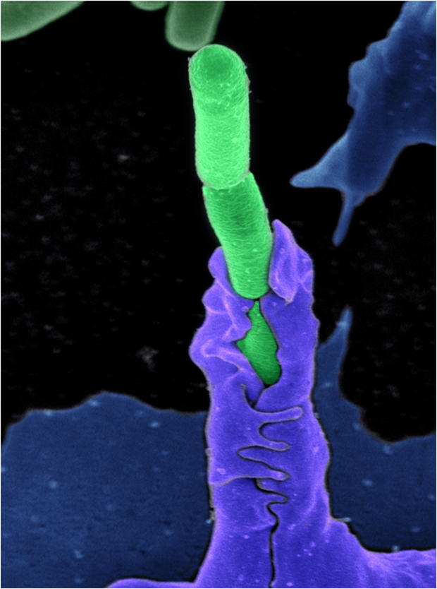
This photo from the exhibit “Life: Magnified” shows the mighty immune system (purple) gulping up a single anthrax bacterium. Anthrax bacteria live in soil and the spores can stay dormant for centuries. When they’re eaten or inhaled the bacteria activatea and rapidly increases in number.
Skin cancer cells
Squamous cell carcinoma is the second most common type of skin cancer. This image shows uncontrolled growth of the cells in a mouse.
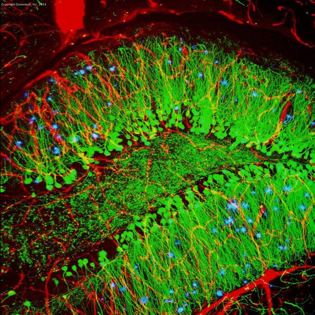
Alzheimer's disease plaque formations
The brain of this lab mouse demonstrates structures of blood vessels (red) and nerve cells (green), but also abnormal proteins proliferating throughout. The blue clumps in this image are plaque buildups, which is a hallmark of Alzheimer’s disease.
Mice have nearly identical genomes to humans and are therefore used in labs to study both genetic and environmental factors that trigger the illness.
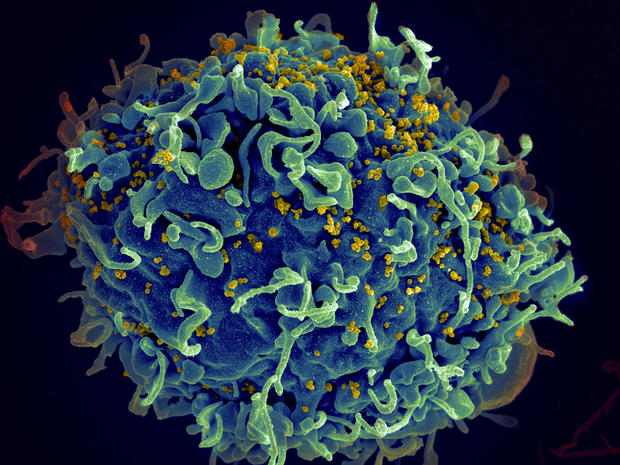
HIV infecting a human cell
This photo from the exhibit “Life: Magnified” shows the HIV virus (yellow) attacking a human T cell. This type of human cell is crucial for a well-functioning immune system to protect the body from bacteria and viruses.
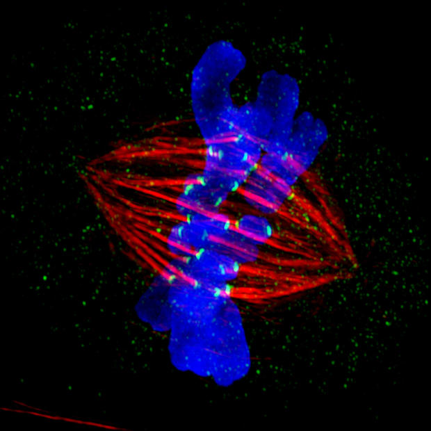
Chromosomes prepare for cell division
Two copies of the each chromosome (blue) are lined up next to each other. The protein strands (red) will soon pull apart these twins and drag them to opposite sides of the cell. The cell will then split to form two daughter cells. If all goes well, each will have a single complete set of chromosomes.
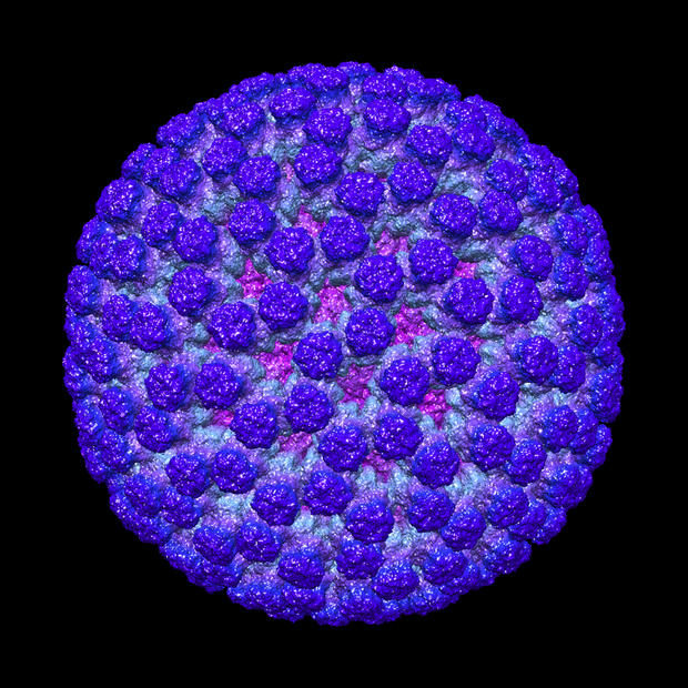
Rotavirus in 3D
This image is a magnification of the rotavirus by about 50,000 time. The rotavirus infects humans as well as animals and causes severe diarrhea in children, though a vaccine is now available in the U.S.
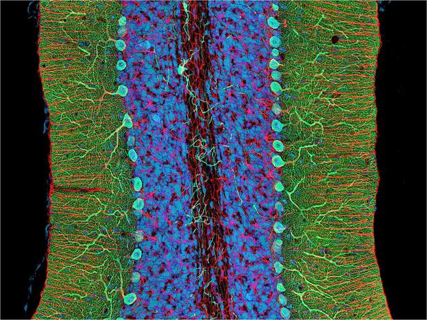
Mouse’s cerebellum
When you walk to work, scramble an egg or sign a check, your cerebellum -- the locomotion control of your brain -- is hard at work. The cerebellum is found at the base of your brain near the spine.
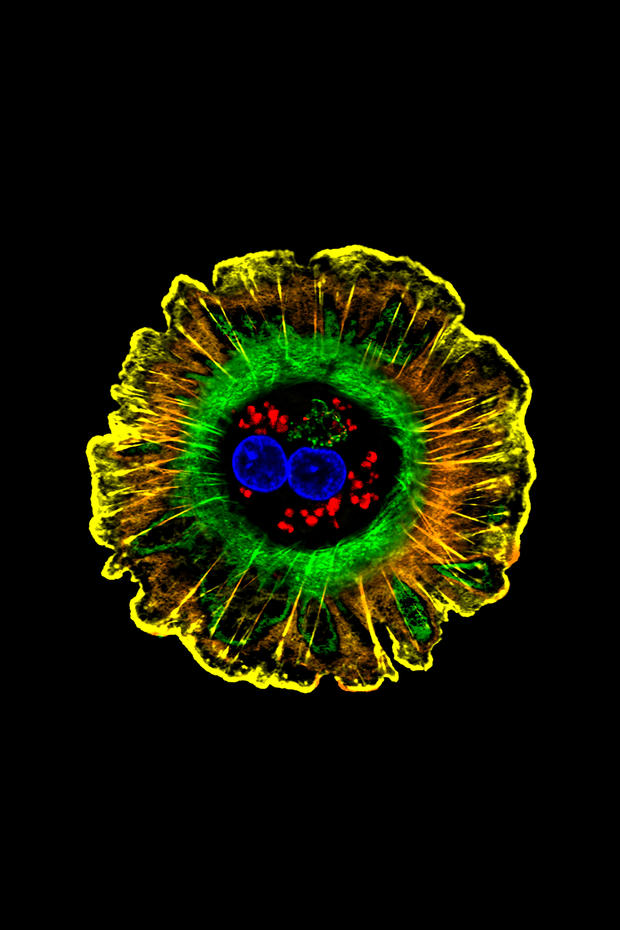
Human liver cell (hepatocyte)
The majority of the liver is made up of hepatocyte. They are crucial for building proteins, producing bile needed for digestion of fats and processing chemicals in the body such as hormones and foreign substances like medicines and alcohol.
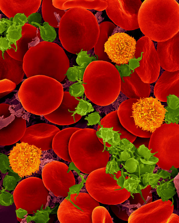
Blood cells
Nearly half of our blood is composed of red blood cells, which deliver oxygen to our tissues. T cells (orange) are an essential part of the immune system. Platelets (green), the smallest blood cells, clump together into clots to stanch bleeding after an injury.
Sound-sensing cells in the ear
Believe it or not, tiny hairs inside your inner ear are integral to your ability to hear. When sound reaches the ear, the hairs bend and the cells send signals to the brain with their movement. Too much sound can cause these tiny hairs to bend until they actually break, resulting in hearing loss.
