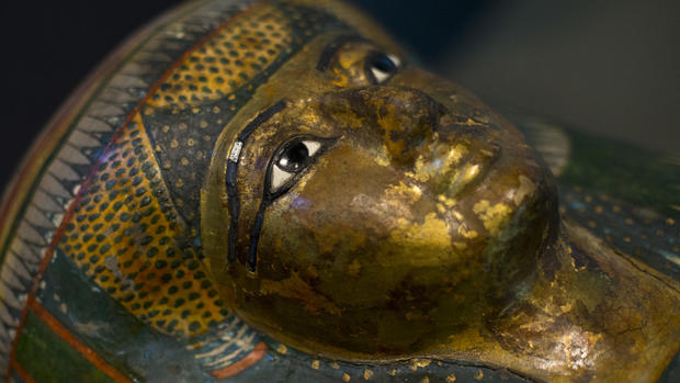
Technology helps unlock secrets of mummies
Doctors have become accustomed to using magnetic resonance imaging or CT scans to assess a whole range of ailments in living human beings. Now, scientists are increasingly using the same technology to unlock the secrets of dead - in many cases the mummified remains of humans dating back thousands of years.
In an entire issue of The Anatomical Record dedicated to mummies, researchers describe how CT (computed tomography) scans - which use X-rays of virtual cross-sections of the body to create three-dimensional images - have become one of the leading diagnostic tools for the in-depth analysis of ancient mummies. Coupled with that has been the use of terahertz imaging or MRIs to give researchers a a more detailed look at mummified tissues.
"Noninvasive imaging of ancient tissues is of increasing interest in paleopathological studies, with conventional X-ray and computed tomography currently considered the diagnostic gold standard," Frank J. Rühli, an author on several papers who is part of the Swiss Mummy Project at the University of Zurich's Centre for Evolutionary Medicine, wrote. Using novel MRI techniques, he added, will broaden available methods for "noninvasive studies on ancient corpses, whether they have wet or dry soft tissue, or bone."
Researchers in the past year have used the technology to determine that one mummy was a girl who lived more than 500 years ago in Peru. In another case, they used a CT scanner to determine that an Egyptian mummy still had her lungs - unusual since the organs were often removed before burial - and that she had an array of small objects around her head that might have been a headdress or shroud. And in still another example, they were able to use it to disprove a theory that King Tut suffered from a painful inflammatory disorder called ankylosing spondylitis.
In a case study highlighted in the journal, Samuel Marquez of the SUNY Downstate Medical Center and his colleagues used data gleaned from three CT scans of Egyptian mummies to explore a hypothesis that paranasal sinus size was related to the environment. The data from the mummies helped strengthen the case that, indeed, sinus size was related to climatic and temperature conditions.
And in what's undoubtedly one of the strangest cases ever to use such technology, scientists and hospital staff at a Dutch hospital used a CT scanner to determine that the mummified remains of a monk were contained inside a 1,000-year-old Buddha statue. The monk had evidently practiced the ancient tradition of self-mummification, in which Buddhist ascetics would starve themselves for years and essentially meditate themselves to death.
Along with the technological advances, the journal also features a literal who's who of mummies around the world - documenting examples from ancient Egypt to those found preserved in Danish bogs to the ice mummies of the Inuit peoples of Greenland. There are also studies detailing the mummies of Papua New Guinea and Peru.
Researchers discussed how studying the conditions and ailments of mummies could prove a boon to modern medicine - prompting some to suggest the time has come to develop a code of ethics for the handling of these specimens.
"Ancient mummies are an especially rich source of information about past human biological conditions as they originated worldwide and from different cultures and time periods," Janet Monge, of the University of Pennsylvania Museum of Archaeology and Anthropology, wrote, along with Rühli, in one of the articles.
"Mummies are likely to yield, for example, more ancient DNA than skeletal remains. The examples of contributions made by mummy studies to our medical knowledge are increasing," they wrote. "This is true particularly to the study of infectious diseases. Naturally and artificially mummified human remains have also been the source for genomic analyses of the adaptive evolution for example of Mycobacterium strains or with more recent mummified samples of the analyses of the Spanish flu."
